
Photo / Image Credits
Some of the great looking images seen on other pages of this website are the creations of outstanding artists & photographers who are listed below.
Home page -
Banner image; by Torsten Wittmann, Scripps Research Institute . Two interphase cells with immunofluorescence labeling of actin filaments (purple), microtubules (yellow), and nuclei (green). This image won first place in the Nikon 2003 Small World photo competition. Source: http://cellimagelibrary.org/images/240 .
"How Can We Help? Panel: Gel Electrophoresis Image - By Mnolf (Photo taken in Innsbruck, Austria) [ GFDL [ http://www.gnu.org/copyleft/fdl.html ], CC-BY-SA-3.0 [http://creativecommons.org/licenses/by-sa/3.0/ ], or CC-BY-SA-2.0 [ http://creativecommons.org/licenses/by-sa/2.0], via Wikimedia Commons.
News & Perspectives Panel - by Gary Halvorson, Oregon State Archives [Attribution], via Wikimedia Commons. Image Source: http://upload.wikimedia.org/wikipedia/commons/5/51/Linoleum_Mural_(Clackamas_County%2C_Oregon_scenic_images)_(clacDA0085).jpg .
_(clacDA0085).jpg)
Scientific & Disease Expertise Panel: Scanning electron micrograph of HIV-1 budding (in green) from cultured lymphocyte. This image has been colored to highlight important features; Photo Credit: C. Goldsmith; Image comes from the Centers for Disease Control and Prevention's Public Health Image Library (PHIL [http://phil.cdc.gov/phil/home.asp]), with identification number #10000. Source: [ http://commons.wikimedia.org/wiki/File%3AHIV-budding-Color.jpg ].
Expertise Main page -
Scientific panel: A 3D model of an Immunoglobulin molecule, showing heavy chains in blue and light chains in green. By the National Library of Medicine, Public Domain, Source .
Industry panel: Structure of well known anti-cancer antibody therapeuric, Herceptin, By RedAndr (self-made, rendered by PyMol) [GFDL, CC-BY-SA-3.0, CC-BY-SA-2.0, or CC-BY-SA-2.5-2.0-1.0], via Wikimedia Commons, Source .
Disease panel - Scanning electron micrograph of HIV-1 budding (in green) from cultured lymphocyte. This image has been colored to highlight important features; Photo Credit: C. Goldsmith; Image comes from the Centers for Disease Control and Prevention's Public Health Image Library (PHIL), with identification number #10000. Source .
Expertise Industry page -
Research panel: Overlay of Strips of spectra of HNCACB and CBCA(CO)NH multidimensional NMR experiments used in determining the 3D structure of proteins in solution. By Axb3 (Own work) [ CC-BY-SA-3.0 ; http://creativecommons.org/licenses/by-sa/3.0/ ], via Wikimedia Commons, Source: [ http://upload.wikimedia.org/wikipedia/commons/a/a6/NMR_sequential_assignment_strips.JPG ].
Development panel: A SDS-PAGE gel from an activity-based proteomics study using probes with different fluorophores in the same lane to simultaneously profile differences in enzyme activities. By Roadnottaken at the English language Wikipedia [GFDL [ http://www.gnu.org/copyleft/fdl.html ], CC-BY-SA-3.0 [ http://creativecommons.org/licenses/by-sa/3.0/ ]], via Wikimedia Commons, Source: http://upload.wikimedia.org/wikipedia/commons/1/18/Gel-abpp_eg.png .
Allied Activities Panel: Working Together Teamwork Puzzle Concept by lumaxart (Working Together Teamwork Puzzle Concept) [CC-BY-SA-2.0; http://creativecommons.org/licenses/by-sa/2.0 ], via Wikimedia Commons, Source: http://upload.wikimedia.org/wikipedia/commons/b/ba/Working_Together_Teamwork_Puzzle_Concept.jpg .
Expertise - Disease page -
Inflammation panel: A segmented polymorphonuclear neutrophil is on the left and, on the right and below, is a eosinophil leucocyte. For comparison the background red blood cell have a diameter of 7-8 micrometers. The picture was taken with a Nikon Eclipse 600 microscope, magnification was 1000x. By Davidcsaba [Dr. David Csaba L.] [Public domain], via Wikimedia Commons, Source: http://upload.wikimedia.org/wikipedia/commons/7/75/Eosinophil_and_polymorphonuclear_neutrophil.jpg .
Cancer panel: [Part of a six-step sequence of the death of a cancer cell. A cancer cell has migrated through the holes of a matrix coated membrane from the top to the bottom, simulating natural migration of a invading cancer cell between, and sometimes through, the vascular endothelium. Notice the spikes or pseudopodia that are characteristic of an invading cancer cell (1). A buffy coat containing red blood cells, lymphocytes and macrophages is added to the bottom of the membrane. A group of macrophages identify the cancer cell as foreign matter and start to stick to the cancer cell, which still has its spikes (2). Shown: Macrophages begin to fuse with, and inject its toxins into, the cancer cell. The cell starts rounding up and loses its spikes (3). As the macrophage cell becomes smooth (4). The cancer cell appears lumpy in the last stage before it dies. These lumps are actually the macrophages fused within the cancer cell (5). The cancer cell then loses its morphology, shrinks up and dies (6). Photo magnification: 3: x8,000 Type: B & W print By Susan Arnold (photographer) [Public domain], via Wikimedia Commons, Source: http://upload.wikimedia.org/wikipedia/commons/5/56/Macs_killing_cancer_cell.jpg .
Infectious Disease panel - S. aureus bacteria escaping destruction by human white blood cells. By =(Credit: NIAID/RML) [Public domain], via Wikimedia Commons, Source: http://upload.wikimedia.org/wikipedia/commons/a/a6/Staphylococcus_aureus_bacteria_escape.jpg .
Expertise - Scientific page -
Glycobiology panel: By Kosi Gramatikoff, User:Stannered (en:Image:Glicoprotein.jpg) [Public domain], via Wikimedia Commons, Source: http://upload.wikimedia.org/wikipedia/commons/e/ed/Glicoprotein.svg .
Structural Biology panel: Three-dimensional model of the whole dodecameric enzyme Calcium/calmodulin kinase II. The image was generated in VMD, using the coordinates provided in Rosenberg et al (2005). Cell 123:765-767. By Lenov (Own work) [GFDL ( http://www.gnu.org/copyleft/fdl.html ), CC-BY-SA-3.0 ( http://creativecommons.org/licenses/by-sa/3.0/ )], via Wikimedia Commons; Source: http://upload.wikimedia.org/wikipedia/commons/0/09/CaMKII-dodecameric.png .
Immunology panel: Neutrophil engulfing anthrax bacteria. By Volker Brinkmann [CC-BY-2.5 ( http://creativecommons.org/licenses/by/2.5 )], via Wikimedia Commons Source: http://commons.wikimedia.org/wiki/File:Neutrophil_with_anthrax.jpg .
Services Offered page -
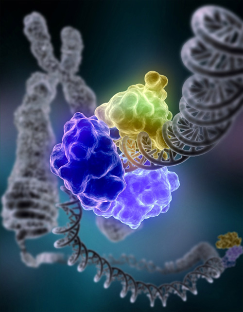
Banner image by courtesy of Tom Ellenberger, Washington University School of Medicine in St. Louis. [Public domain], via Wikimedia Commons; Source: [ http://upload.wikimedia.org/wikipedia/commons/4/46/DNA_Repair.jpg ]. The enzyme DNA ligase I is shown repairing chromosomal damage. The three visable protein structures are: 1) The DNA binding domain (DBD) which is bound to the DNA minor groove both upstream and downstream of the damaged area, 2) The OB-fold domain (OBD) unwinds the DNA slightly over a span of six base pairs and is generally involved in nucleic acid binding, and 3) The Adenylation domain (AdD) contains enzymatically active residues that join the broken nucleotides together by catalyzing the formation of a phosphodiester bond between a phosphate and hydroxyl group.
News With Views page -
Banner image: " DNA at ICSB 2008" By Duncan Hull [dullhunk], License - Attribution: http://creativecommons.org/licenses/by/3.0/ . Some rights reserved by dullhunk, Source: http://www.flickr.com/photos/dullhunk/2225574423/sizes/o/in/photostream/ .
About Us page -
Image by Peter Kwong [ http://www3.niaid.nih.gov/labs/aboutlabs/VRC/structuralBiologyLaboratory/kwong.htm ], [Public domain], via Wikimedia Commons; Source: http://upload.wikimedia.org/wikipedia/commons/9/90/Hiv_ligand_receptor_binding.jpg . The HIV gp120 envelope glycoprotein binds sequentially to the cellular receptors CD4 and a member of the chemokine receptor family (CXCR4 or CCR5) to initiate virus cell entry; Kwong et al., Nature, 420, 678-682 (12 December 2002). The antibody-protein binding to the CD4 receptor attempts to block the chemokine-receptor binding, thus prevent HIV-1 virus cell entry. Both gp120 and CD4 are glycoproteins Kwong et al., Nature 393, 648-659 (18 June 1998).
Colleagues page -
Banner image By Christoph Bock (Max Planck Institute for Informatics) (Own work) [CC-BY-SA-3.0; http://creativecommons.org/licenses/by-sa/3.0/ ], via Wikimedia Commons; Source: http://upload.wikimedia.org/wikipedia/commons/8/80/DNA_methylation.jpg . A DNA molecule that is methylated on both strands on the center cytosine. DNA methylation plays an important role for epigenetic gene regulation in development and cancer. [Details: The picture shows the crystal structure of a short DNA helix with sequence "accgcCGgcgcc", which is methylated on both strands at the center cytosine.
Contact Us page -
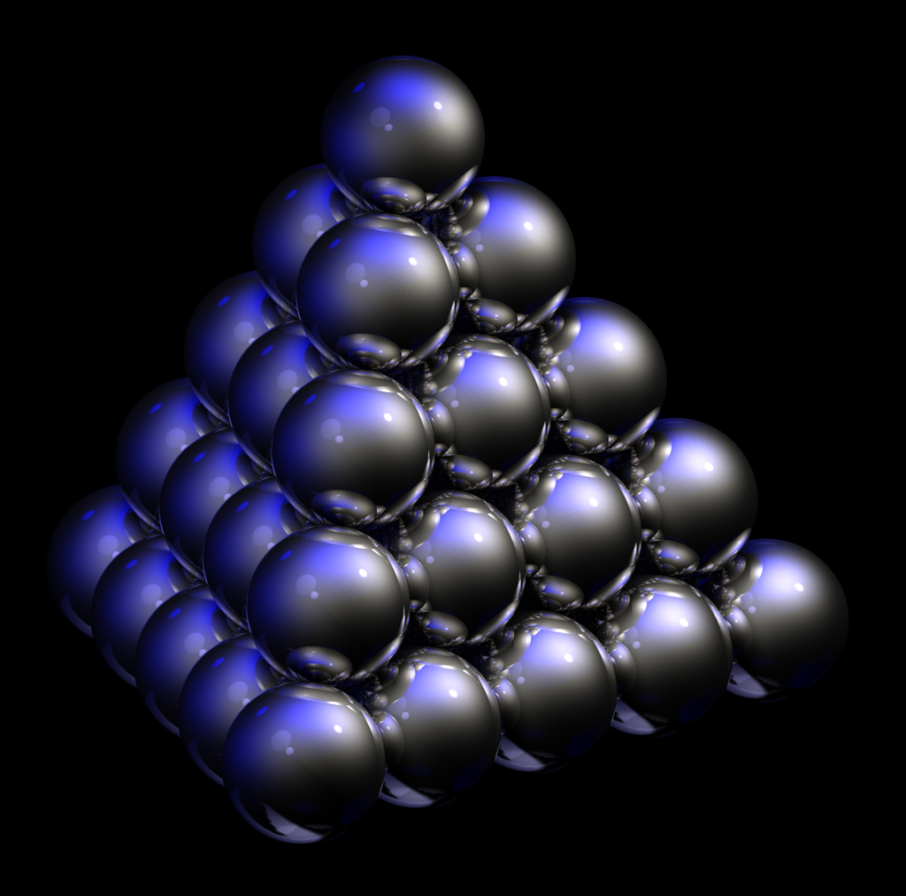
Banner image: "Close-packed spheres"; Attribution: [GFDL [ http://www.gnu.org/copyleft/fdl.html ], CC-BY-SA-3.0 ( http://creativecommons.org/licenses/by-sa/3.0/ )], via Wikimedia Commons; Source: http://upload.wikimedia.org/wikipedia/commons/8/8e/Close-packed_spheres.jpg .
LinkedIn Logo - By LinkedIn, User:ZyMOS [Public domain], via Wikimedia Commons
Skype Logo - By User:ZyMOS [Public domain], via Wikimedia Commons
Infectious Disease News With Views page -
Banner image: MRSA (yellow) being ingested by neutrophil (purplish blue). Credit: NIAID; Attribution: By NIAID/NIH (NIAID Flickr's photostream) [Public domain], via Wikimedia Commons; Source: http://commons.wikimedia.org/wiki/File%3AMethicillin-resistant_Staphylococcus_aureus_Bacteria.jpg
World AIDS Day Image - Ribbon: By de:User:ChristianHeldt [Public domain], via Wikimedia Commons. Source: http://upload.wikimedia.org/wikipedia/commons/e/e6/World_Aids_Day_Ribbon.png
Cancer News With Views page -
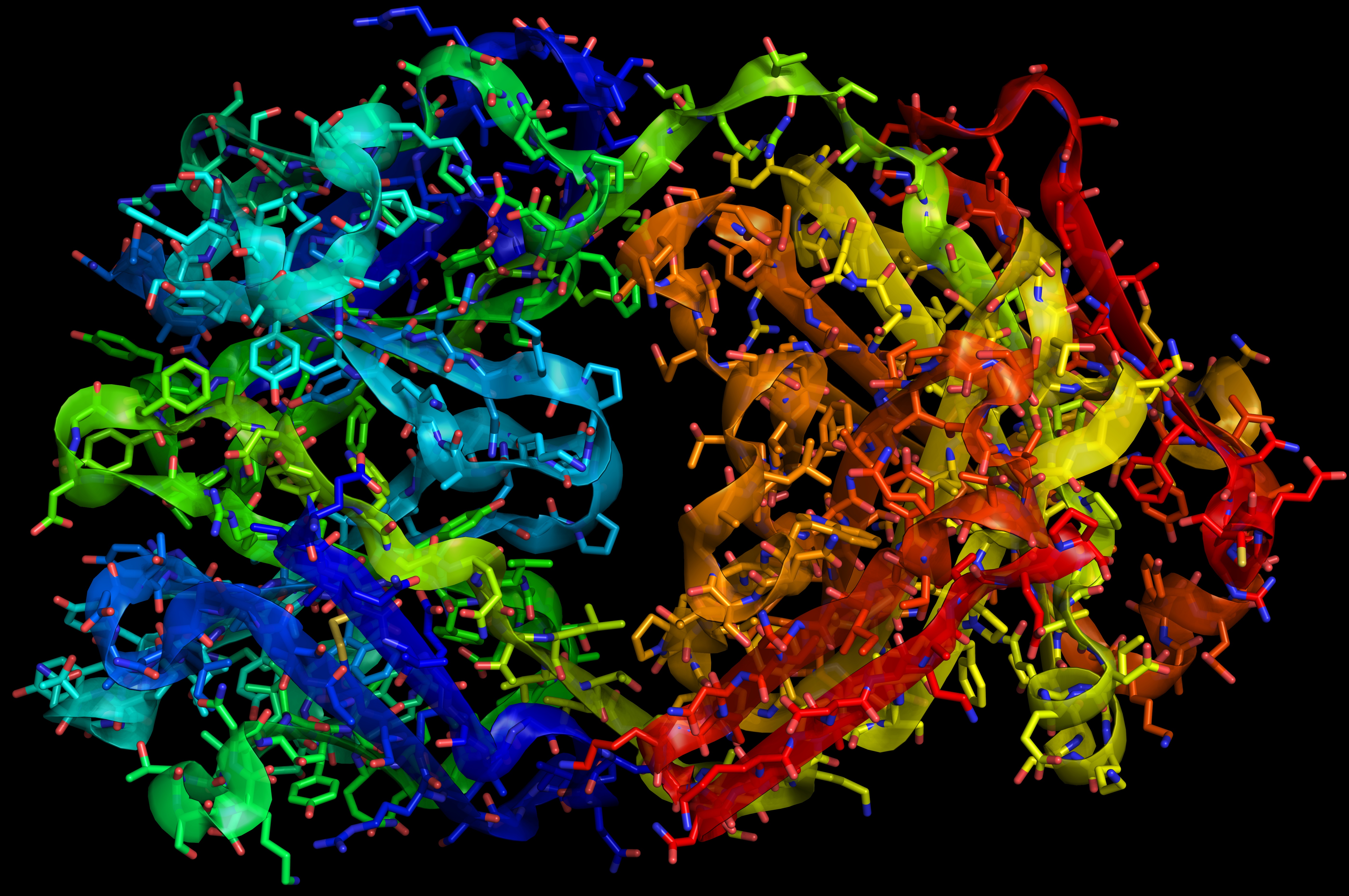
Banner image: Partial molecular structure of the anti-cancer monoclonal antibody therapeutic Heceptin. Attribution: By RedAndr (self-made, rendered by PyMol) [GFDL (<span><a href="http://www.gnu.org/copyleft/fdl.html" class="smarterwiki-linkify">http://www.gnu.org/copyleft/fdl.html</a></span>), CC-BY-SA-3.0 (<span><a href="http://creativecommons.org/licenses/by-sa/3.0/" class="smarterwiki-linkify">http://creativecommons.org/licenses/by-sa/3.0/</a></span>) or CC-BY-SA-2.5-2.0-1.0 (<span><a href="http://creativecommons.org/licenses/by-sa/2.5-2.0-1.0" class="smarterwiki-linkify">http://creativecommons.org/licenses/by-sa/2.5-2.0-1.0</a></span>)], via Wikimedia Commons; Source: http://upload.wikimedia.org/wikipedia/commons/0/0a/HerceptinFab.jpg . Breast Cancer Awareness Month banner image: "Pink ribbon chocolates" By wishuponacupcake [CC-BY-2.0, via Wikimedia Commons]; source: http://upload.wikimedia.org/wikipedia/commons/6/67/Pink_Ribbon_chocolates.jpg.
Inflammation News With Views page -
Banner image: Pollen from a variety of common plants: sunflower (Helianthus annuus), morning glory Ipomoea purpurea, hollyhock (Sildalcea malviflora), lily (Lilium auratum), primrose (Oenothera fruticosa) and castor bean (Ricinus communis). Attribution: The colorization of this illustration was made by Medium69 Please credit this : William Crochot Website : http://www.science-et-vie.net. Credit: Dartmouth Electron Microscope Facility, Dartmouth College [Public domain], via Wikimedia Commons. Source: http://upload.wikimedia.org/wikipedia/commons/2/2a/Misc_pollen_colorized.jpg
Other (Misc.) Science News With Views page -
Banner image: Collection of various fluorescent minerals under ultraviolet UV-A, UV-B and UV-C light. Chemicals in the rocks absorb the ultraviolet light and emit visible light of various colors. Credit: By Hannes Grobe (Hgrobe 06:16, 26 April 2006 (UTC)) (Own work) [CC-BY-SA-2.5], via Wikimedia Commons. Source: http://upload.wikimedia.org/wikipedia/commons/1/12/Fluorescent_minerals_hg.jpg .
Glycobiology News With Views page -
Banner image: Multiple fluorescence 2PE imaging. 2PE multiple fluorescence image from a 16 μm cryostat section of mouse intestine stained with a combination of fluorescent stains (F-24631, Molecular Probes). Alexa Fluor 350 wheat germ agglutinin, a blue-fluorescent lectin, was used to stain the mucus of goblet cells. The filamentous actin prevalent in the brush border was stained with red-fluorescent Alexa Flu or 568 phalloidin. Finally, the nuclei were stained with SYTOX ® Green nucleic acid stain. Imaging has been performed at 780 nm, 100 x 1.4 NA Leica objective, using a Chameleon XR ultrafast Ti-Sapphire laser (Coherent Inc., USA) coupled at LAMBS-MicroScoBio with a Spectral Confocal Laser Scanning Microscope, Leica SP2-AOBS. Credit: By Alberto Diaspro, Paolo Bianchini, Giuseppe Vicidomini, Mario Faretta, Paola Ramoino and Cesare Usai [CC-BY-2.0], via Wikimedia Commons. Image source: http://upload.wikimedia.org/wikipedia/commons/3/36/MultiPhotonExcitation-Fig10-doi10.1186slash1475-925X-5-36.JPEG. Please see the author's paper at http://www.biomedical-engineering-online.com/content/5/1/36 .
Immunology News With Views page -
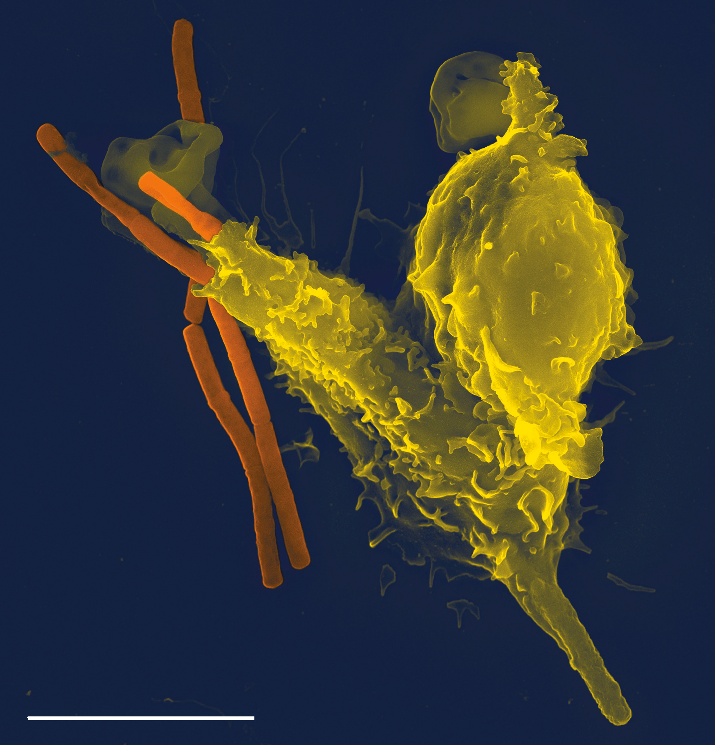
Banner image: Neutrophil (yellow) engulfing rod-shaped Bacillus anthracis (anthrax) bacteria (orange), taken with a Leo 1550 scanning electron microscope. Neutrophils are the most abundant white blood cell and the first line of defense against invading microbes. A larger image version was the cover for PLoS Pathogens 1 (3) in November of 2005. Credit: By Volker Brinkmann [CC-BY-2.5 ], via Wikimedia Commons. Source: http://upload.wikimedia.org/wikipedia/commons/f/f2/Neutrophil_with_anthrax_copy.jpg .
Vaccine News With Views page -
Banner images: "Wellbee" Polio Poster - Photo Credit: Content Providers(s): CDC/ Mary Hilpertshauser [Public domain], via Wikimedia Commons. This 1963 poster featured CDC’s national symbol of public health, the "Wellbee", who was depicted here encouraging the public to receive an oral polio vaccine. The CDC used the Wellbee in its comprehensive marketing campaign that used newspapers, posters, leaflets, radio and television, as well as personal appearances at public health events. Wellbee’s first assignment was to sponsor Sabin Type-II oral polio vaccine campaigns across the United States. Later, Wellbee’s character was incorporated into other health promotion campaigns including diphtheria and tetanus immunizations, hand-washing, physical fitness, and injury prevention. This artifact can be found in the Global Health Odyssey, which is the CDC’s museum featuring many various public health-related artifacts. Source: http://upload.wikimedia.org/wikipedia/commons/b/b8/Polio_vaccine_poster.jpg; "Wellbee" Get a Booster poster - Photo Credit: Content Providers(s): CDC/ Mary Hilpertshauser [Public domain], via Wikimedia Commons. This 1964 poster featured what at that time, was CDC’s national symbol of public health, the “Wellbee”, who here was reminding the public to get a booster vaccination. Source: http://upload.wikimedia.org/wikipedia/commons/8/85/BoosterWellbeeSpace7220.jpg; Diphtheria Vaccination Poster - This artistic work created by the United Kingdom Government is in the public domain. Source: http://upload.wikimedia.org/wikipedia/commons/8/8f/Diphtheria_vaccination_poster.jpg .
Adobe and Adobe Muse are either registered trademarks or trademarks of Adobe Systems Incorporated in the United States and/or other countries.
Apple, the Apple logo, and Mac are trademarks of Apple Inc., registered in the U.S. and other countries. The Made on a Mac Badge is a trademark of Apple Inc., used with permission.
© 2012-2014 Acton Biotech Consulting - See Image Credits page for attribution and license conditions for non-original images/media.
Important DISCLAIMER - This is a science & technology website and not a medical treatment or diagnostic site. No information contained within this site is a substitute for advice or direction given by qualified medical professionals, nor is it intended to inform patients regarding treatment options or disease diagnosis/prognosis. As always, individuals should consult their own medical team about issues concerning their health and well being.